
Astounding new research has revealed malignant cancer in dinosaur bones.

Scientists from the Royal Ontario Museum (ROM) and McMaster University made the discovery and diagnosis of an aggressive malignant bone cancer — an osteosarcoma — for the first time ever in a dinosaur.

Mark Crowther, Professor of Pathology and Molecular Medicine said:
“Diagnosis of aggressive cancer like this in dinosaurs has been elusive and requires medical expertise and multiple levels of analysis to properly identify.”
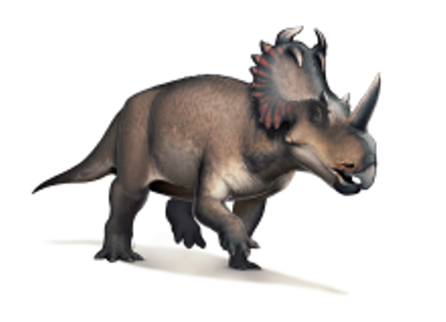
The cancerous bone in question is the fibula (lower leg bone) from Centrosaurus apertus, a horned dinosaur that lived 76 to 77 million years ago.
Originally discovered in Dinosaur Provincial Park in Alberta in 1989, the badly malformed end of the fossil was originally thought to represent a healing fracture.
After carefully examining, documenting, and casting the bone, the team performed high-resolution computed tomography (CT) scans. They then thin-sectioned the fossil bone and examined it under a microscope to assess it at the bone-cellular level. Powerful three-dimensional CT reconstruction tools were used to visualize the progression of the cancer through the bone. Using this rigorous process, the investigators reached a diagnosis of osteosarcoma.
To confirm this diagnosis, they then compared the fossil to a normal fibula from a dinosaur of the same species, as well as to a human fibula with a confirmed case of osteosarcoma. The fossil specimen is from an adult dinosaur with an advanced stage of cancer that may have invaded other body systems. Yet it was found in a massive bone bed, suggesting it died as part of a large herd of Centrosaurus struck down by a flood.

Dr. David Evans, James and Louise Temerty Endowed Chair of Vertebrate Palaeontology from the ROM, said:
“The shin bone shows aggressive cancer at an advanced stage. The cancer would have had crippling effects on the individual and made it very vulnerable to the formidable tyrannosaur predators of the time.
“The fact that this plant-eating dinosaur lived in a large, protective herd may have allowed it to survive longer than it normally would have with such a devastating disease.”
The results were published in the medical journal The Lancet Oncology.
This 3D animation, reconstructed based on high-resolution X-ray computed tomography (HRXCT) scans, shows the main tumor mass (yellow) at the top of the fibula as well as the progression of the tumour into the other end of the bone. The malignant tumour is in yellow; the normal bone is in gray, and red delineates the medullary cavity.


Leave a Reply