
Every gardener knows how important the earthworm is in producing healthy soils: burrowing, pulling down leaves, aerating the soil- and as a food source themselves for our blackbirds.

Now a team of researchers around Benedikt Geier from the Max Planck Institute for Marine Microbiology (MPIMM) in Bremen, Germany, has succeeded in imaging the exciting variety of interactions that take place inside the earthworm.
This allows us to observe them in a completely new light.
Researcher Benedikt Geier explains:
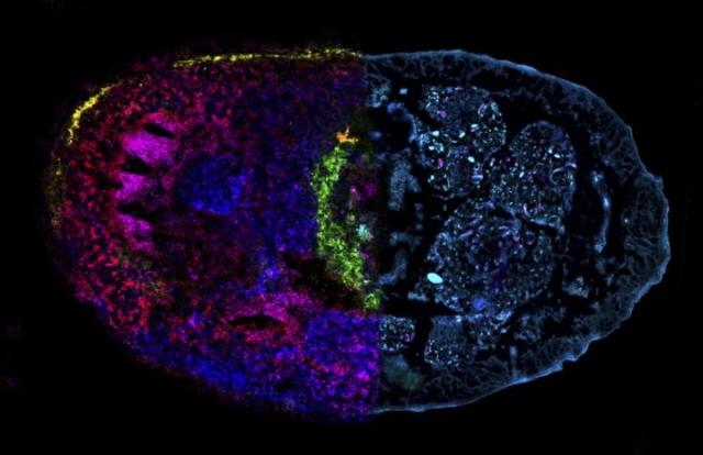
“In our study, we introduce chemo-histo-tomography, a special three-dimensional imaging approach of the chemistry and anatomy of millimeter-sized animals and their parasites at a cellular level. This method offers a new strategy to visualize the most fundamental processes – metabolic interactions – in small animal symbioses. “

Using the combination of different in situ imaging techniques through CHEMHIST (Chemo-histo-tomography), the researchers at the MPIMM in Bremen uncovered metabolites – products of the metabolism – in the earthworm that could shed light on how it chemically defends itself against parasites and how these, in turn, protect themselves against the earthworm’s immune response.
The team were able to create a 3D atlas of its chemical and physical interactions with the microorganisms naturally occurring inside its tissues. Extending correlative chemical imaging into 3D approaches can be crucial for capturing the distribution of metabolites involved in symbiotic interactions and thus show how chemical signals from microbes possibly affect crucial processes in their host. The method can be used in many ways: The research team around Liebeke and Geier is currently applying it to deep-sea mussels.

The results of the study were made possible by an interdisciplinary collaboration that brought together scientists from the fields of microbiology, zoology, chemistry and physics. It quickly became clear that visualization such as the correlative 3D atlas of the worm facilitate science communication of the data.
You can read more about the research here: Connecting structure and function from organisms to molecules in small-animal symbioses through chemo-histo-tomography


Leave a Reply